Protecting the cultural heritage of ancient bone artifacts is now possible. Near-infrared hyperspectral imaging and radiocarbon dating together to make the invisible visible
Developed by scientists from the University of Bologna and the University of Genoa, the new technology makes it possible to map at high resolution the presence of collagen, the protein that is essential for carrying out radiocarbon dating. It will then be possible to strategically sample artifacts, identifying suitable fragments and areas for analysis
An innovative method developed by an Italian team is emerging that will revolutionize the field of archaeology and radiocarbon dating and protect our cultural heritage. The researchers have used it with surprising results on archaeological bones, making the ‘invisible’ visible.
This important achievement - published in the journal Communications Chemistry of the Nature group - is the result of extensive research work coordinated by Professor Sahra Talamo, in which experts in the field of analytical chemistry from the University of Bologna and the University of Genoa collaborated.
The group has developed a new technique for analyzing archaeological bones that, for the first time, makes it possible to quantify and map at high resolution the presence of collagen, the invisible protein that is essential for making radiocarbon dates and thus obtaining new information on human evolution.
"Our results will offer significant advances for the study of human evolution," says Talamo coauthor of the study and director of the Radiocarbon dating lab BRAVHO at the University of Bologna. "As we will be able to minimise the destruction of valuable bone material, which is under the protection and enhancement of European cultural heritage and thus allow us to contextualise the valuable object by providing an accurate calendar age."
Many of the rarest prehistoric bones found by archaeologists are enormously precious and are considered to be part of our cultural and historical patrimony. Bones can provide a great deal of information about ancient populations’ lives: what they ate, their reproductive habits, their diseases and the migrations they undertook. However, bones cannot give us all the information we so covet. Their potential to convey information is limited by how much collagen is preserved in them.
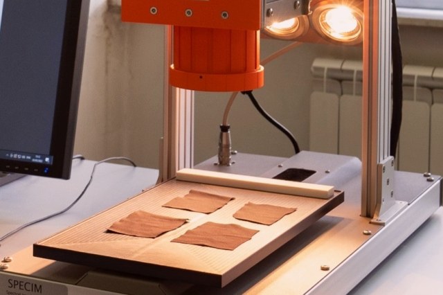
In order to combine the need to preserve the integrity of the artifacts as much as possible with the need to carry out radiocarbon analyses, the researchers therefore developed an innovative method that, thanks to a camera coupled with near-infrared, allows them to detect the average collagen content in the observed samples.
"We used imaging technology to quantify the presence of collagen in bone samples in a non-destructive way to select the most suitable samples (or sample regions) to be submitted to radiocarbon dating analysis," says Cristina Malegori, first author of the article and researcher at Genoa University, Department of Pharmacy. "Near-infrared hyperspectral imaging (HSI) was used along with a chemometric model to create chemical images of the distribution of collagen in ancient bones. This model quantifies the collagen at every pixel and thus provides a chemical mapping of collagen content."
It is extremely difficult, costly, and time-consuming to analyze all the bones present at one archaeological site for collagen preservation, most importantly, it would result in the destruction of valuable material. In fact, human fossils and bone artifacts are increasingly rarer and more precious over time. Because of the diagenetic alteration of collagen over time, large starting weights of Palaeolithic bones (≥ 500 mg bone material) are necessary to extract sufficient collagen for accelerator mass spectrometry (AMS) 14C dating (minimum 1% yield). Moreover, many of the most precious archaeological bones are too small (< 200 mg of bone material) or too beautiful for sampling. Therefore, obtaining preliminary, non-destructive information about the distribution of collagen on a bone sample is crucial.
It is in this context that the technique described in this study really shines because it allows obtaining information both on the location and on the content of the collagen still present in a bone sample.
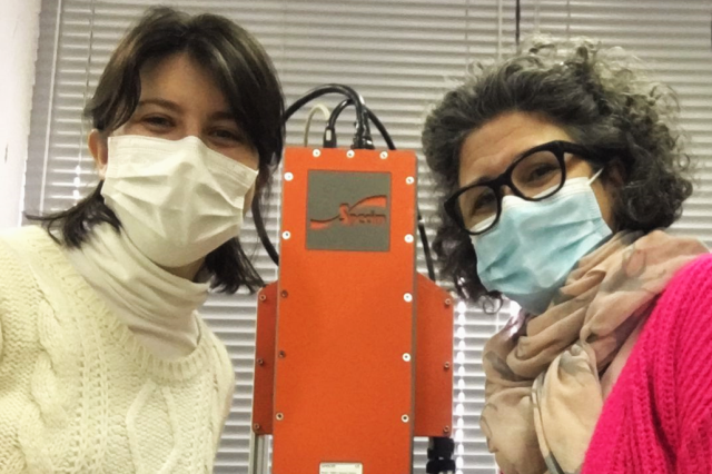
Cristina Malegori (University of Genoa) and Sahra Talamo (University of Bologna)
"The near-infrared hyperspectral imaging camera (NIR-HSI) used in the present study is a line-scan (push-broom) system that acquires chemical images in which, for every pixel, a full spectrum in the 1,000–2,500 nm spectral range (near infrared) is recorded," says Giorgia Sciutto, co-author of the article and professor of environmental and cultural heritage chemistry at the University of Bologna. "NIR-HSI analysis is completely non-destructive. The time required for the analysis of a single bone sample is of few minutes and, therefore, the system can examine many samples in a single day to find those suitable for analysis, saving time and money and the unnecessary waste of valuable material, greatly reducing time, costs and destruction of valuable samples."
This technique is expected to support the selection of samples to be submitted to radiocarbon analysis at many sites where previous attempts have not been possible because of poor preservation.
"This new technique allows not only selecting the best specimens but also choosing the sampling point in the selected ones based on the amount of collagen predicted," says Paolo Oliveri co-author of the paper and professor at the Genoa University, Department of Pharmacy. "This method helps to drastically reduce the number of samples destroyed for 14C analysis, and within the bone, it helps to avoid the selection of areas that may present a quantity of collagen not sufficient for the dating. This increases the preservation of precious archaeological materials."
"The potential of the method proposed in the present study lies in the type and amount of information that the predictive model provides, addressing two fundamental and complementary questions for the characterization of collagen in bones: how much and where," says Cristina Malegori, first author of the article.
Thus, this experimental approach can provide quantitative information related to the average collagen content present in the whole sample submitted for investigation. The examination can be performed not only in small and localized areas (as in single-point analysis), but it can also consider the entire surface of the sample, thus producing a higher and much more significant amount of data. In addition, combining the HSI system with PLS regression allowed, for the first time, on samples of ancient bones, not only to determine the overall collagen content but also to localize it at a high spatial resolution (about 30 um), obtaining quantitative chemical maps.
"As far as radiocarbon is concerned, we could strategically sample bones of high patrimonial value. For example, knowing the precise amount of collagen concentrated in a precise area of the bone allows us to cut only this portion," says Talamo. "Moreover when the prediction of collagen shows that the bone was poorly preserved, we can decide to perform a soft 14C pretreatment to minimize collagen loss during the extraction."
Overall, this innovative and incisive combination of NIR-HSI spectroscopy prescreening and the radiocarbon method provides, for the first time, detailed information about the presence of collagen on archaeological bones, reducing laboratory costs by dating only materials suitable for 14C and increasing the number of archaeological bones that can be preserved, and, therefore, available for future research.


