At the University of Bologna, the First CT Scanner in Italy Dedicated to Research in Human Anatomy
The new Philips Incisive CT scanner was presented at the University of Bologna’s Anatomy Centre: with this unique project, anatomical dissection—one of the oldest scientific practices—merges with cutting-edge digital technologies, generating new knowledge and paving the way for increasingly personalised, safe and effective healthcare and training models
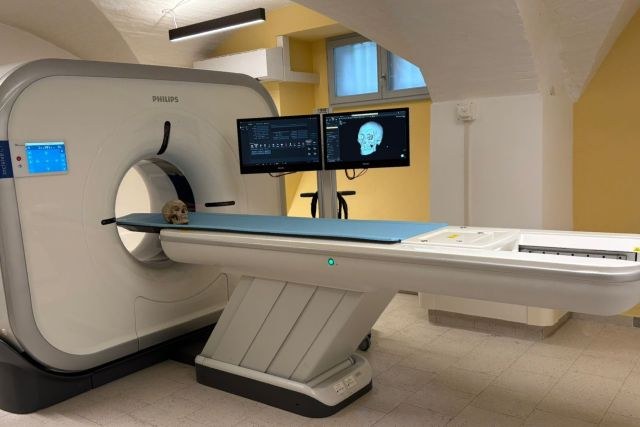
The University of Bologna is inaugurating a new phase in scientific research and medical education with the presentation of the new Philips Incisive CT scanner, installed at the Centre for Clinical, Surgical, Experimental and Molecular Anatomy within the Department of Biomedical and Neuromotor Sciences. Funded through Italy’s NRRP – Mission 6 Health, it is the first CT scanner in the country to be installed at a university institute with exclusive use for research and teaching in human anatomy.
"Thanks to this technology, anatomical images become imaginative tools that can support personalised surgical simulations and high-level training," explains Stefano Ratti, professor of Anatomy at the University of Bologna and the project's lead promoter. "From a donated body come new possibilities for care and knowledge—an act of generosity that drives innovation."
The new CT scanner enables ultra-precise 3D reconstructions of bodies donated to science and received by the University of Bologna’s Anatomy Centre, which is a nationally accredited reference centre recognised by the Ministry of Health. This opens up opportunities for applying the technology in areas such as personalised surgery, 3D modelling for medical training, and precision medicine—placing patients and procedural safety at the centre.
“We are proud to support the research activities of the Anatomy Centre with our CT system, which, thanks to artificial intelligence, offers clear visualisation of complex anatomical structures,” says Valeria Nardella, CT Product Manager at Philips Italy. This is an application beyond the traditional clinical setting that demonstrates how innovation can support research and education in the service of precision medicine and public health.”
“For the first time, we can combine direct observation of the body with advanced virtual modelling,” says Emanuela Marcelli, coordinator of the EDIMES Lab (Biomedical Engineering Laboratory) and professor at the University of Bologna. “This allows us to engage with the human body with unprecedented precision and flexibility, benefitting both research and teaching.”
“In the field of maxillofacial surgery, advanced anatomical imaging is revolutionising our approach to complex reconstructions,” adds Giovanni Badiali, surgeon and professor at the University of Bologna. “Access to realistic 3D models derived from donated bodies allows for safer, more targeted and more effective surgical planning.”
The presentation of the new equipment comes just weeks after the national launch of the campaign to promote body donation to science, held on 15 May in the Aula Magna of Santa Lucia. This marks a symbolic and concrete connection between the ethics of donation and the advancement of scientific research, placing at the centre the priceless value of the generosity of body donors—today more essential than ever for the development of future medicine.