Osteosarcoma is the most frequent malignant bone tumour in children and adolescents. It is an aggressive disease that remains difficult to treat, as currently available therapies are still not sufficiently effective and have not substantially changed over the past decades.
One of the main obstacles is the lack of experimental models capable of faithfully reproducing the complex interaction between tumour cells and their surrounding microenvironment.
The new project led by the Department of Pharmacy and Biotechnology aims to overcome this limitation by creating a true three-dimensional “avatar” of the tumour.
The study will involve postdoctoral researcher Francesca Rossi, supported by a post-doctoral fellowship from AIRC – Italian Association for Cancer Research.
“This project will allow us to recreate the tumour in the laboratory in a much more realistic way than in the past, and to observe how it develops and responds to drugs in an environment that closely resembles what happens in the human body,” explains Dr Rossi. “It is an important step towards a better understanding of the disease and towards identifying new therapeutic strategies for young patients.”
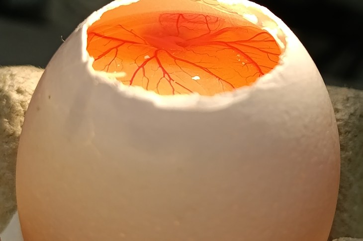
The approach is based on observing the growth of osteosarcoma cells in three-dimensional aggregates that reproduce the architecture and molecular processes of the tumour. These cell aggregates will then be implanted onto the chorioallantoic membrane (CAM) of a chicken embryo – a naturally highly vascularised system that will act as an incubator. This will enable the tumour to develop in a living, vascularised environment, allowing researchers to observe key phenomena such as tumour growth, the formation of new blood vessels, invasion of surrounding tissues and responses to drugs under conditions that are much closer to those experienced by patients.
The use of the CAM is not classified as animal experimentation and provides a low-cost platform that can be readily translated towards clinical applications.
The tumour will be analysed using advanced, extremely high-power X-ray techniques generated by synchrotron light. Their brightness is in fact one hundred thousand billion billion times greater than that of the Sun – a huge difference that makes it possible to study the tumour down to the atomic scale.
The ultimate goal is to develop a predictive experimental system to better understand the mechanisms underlying osteosarcoma, to test new drugs and therapeutic strategies, and to identify the most promising ones in order to pave the way for future precision-medicine approaches.
The two-year post-doctoral fellowship makes it possible to structure the project in several phases, ranging from the development of the experimental model to the biological and structural characterisation of the tumour and, finally, to pharmacological screening.
The research is carried out under the supervision of Professor Emil Malucelli, Professor of Clinical Biochemistry and Clinical Molecular Biology at the University of Bologna, and is part of a national and international network of collaborations involving leading research centres and infrastructures.